
Comparative In-Silico Sequence and Structural Analysis of Catalase from Escherichia coli and Humans
DOI:
https://doi.org/10.33003/fjs-2026-1019-6183Keywords:
In-silico analysis, Catalase, Escherichia coli, Sequence alignment, Oxidative stressAbstract
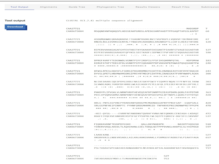
Hydrogen peroxide (H2O2) is a significant reactive oxygen species that can lead to cellular damage, particularly in eukaryotic organisms. Catalase, an enzyme, plays a crucial role in detoxifying H2O2 and sustaining cellular functions. Although substantial research has been conducted on catalase, limited comparative analyses examining the catalase sequences between Escherichia coli and humans, particularly in the context of active sites, exist. This study retrieved and analyzed the amino acid sequences and three-dimensional structures of catalase from both organisms using NCBI and PDB, alongside tools such as Clustal Omega, BLAST, MEGA, and PyMOL. The multiple sequence alignment identified 200 conserved, 122 physiochemically similar, and 163 variable amino acid residues, corresponding to an overall sequence identity of 45.17%. Phylogenetic analysis of a small representative dataset illustrated the evolutionary relationship between E. coli and human catalase. Structural superimposition of the active-site regions yielded a root mean square deviation (RMSD) of 0.295 Å, indicating close structural correspondence in this region despite the divergence in overall sequence and length. Essential functional residues, including active sites, domains, and motifs, were identified and compared. This computational analysis offers preliminary insights into the evolutionary and structural relationships of catalase between the two organisms, which may help direct future experimental work on oxidative-stress-related enzyme function.
References
Baker, A., Lin, C. C., Lett, C., Karpinska, B., Wright, M. H., & Foyer, C. H. (2023). Catalase: A critical node in the regulation of cell fate. Free radical biology & medicine, 199, 56–66. https://doi.org/10.1016/j.freeradbiomed.2023.02.009
Nandi, A., Yan, L. J., Jana, C. K., & Das, N. (2019). Role of Catalase in Oxidative Stress- and Age-Associated Degenerative Diseases. Oxidative medicine and cellular longevity, 2019, 9613090. https://doi.org/10.1155/2019/9613090
Tian, Y., Liu, Y. R., Jin, H. Z., Lin, Q. X., Zhao, W. Y., Song, W. W., Gong, Y. N., Deng, Y. T., Wang, S. S., Wang, K., Tian, L., & Gu, D. N. (2026). Catalase inhibits tumor growth by alleviating oxidative stress and promoting the polarization of tumor-associated macrophage from M2 to M1 phenotype in non-small cell lung cancer. Biology direct, 21(1), 24. https://doi.org/10.1186/s13062-026-00730-6
Glorieux, C. & Calderon, P. (2017). Catalase, a remarkable enzyme: targeting the oldest antioxidant enzyme to find a new cancer treatment approach. Biological Chemistry, 398(10), 1095-1108. https://doi.org/10.1515/hsz-2017-0131
Scholz, A. T., Coppo, L., Nolan, E., Hernandez, M., Wang, X., Mishra, P., Schnell, R., Pató, Z. A., Chen, Y., Dagnell, M., Andor, A., Cheng, Q., & Arnér, E. S. J. (2026). A katE katG double-knockout E. coli strain eliminates the risk of catalase contamination in recombinant proteins. Applied microbiology and biotechnology, 110(1), 136. https://doi.org/10.1007/s00253-026-13820-2
Rasheed, Z. (2024). Therapeutic potentials of catalase: Mechanisms, applications, and future perspectives. International Journal of Health Sciences, 18(2), 1–6. PMID: 38455600.
Olufunmilayo, E. O., Gerke-Duncan, M. B., & Holsinger, R. M. D. (2023). Oxidative stress and antioxidants in neurodegenerative disorders. Antioxidants, 12(2), 517. https://doi.org/10.3390/antiox12020517
Sayers, E. W., Bolton, E. E., Brister, J. R., Canese, K., Chan, J., Comeau, D. C., Connor, R., Funk, F., Murphy, T., Phan, L., Skripchenko, Sherry, S. T. (2022). Database resources of the National Center for Biotechnology Information. Nucleic Acids Research, 50(D1), D20–D26. https://doi.org/10.1093/nar/gkab1112
Shahid, M., Amjad, M., Ashraf, U., Jamal, A., & Wattoo, J. (2021). Computational analysis of catalase from different source organisms. Pakistan Journal of Botany. 54(35). https://doi.org/10.30848/PJB2022-1
Sievers, F., & Higgins, D. G. (2018). Clustal Omega for making accurate alignments of many protein sequences. Protein science : a publication of the Protein Society, 27(1), 135–145. https://doi.org/10.1002/pro.3290
Timsina, R., Gora, R. A., & Ferguson, D. J., Jr (2025). Proteomic and metabolomic analysis reveals new insights into quaternary amine metabolism in Citrobacter amalonaticus CJ25. mSphere, 10(9), e0042125. https://doi.org/10.1128/msphere.00421-25
Jones, D. (2021). Student Tutorial for PyMOL: for educational use Visualizing proteins using molecular graphics. 10.13140/RG.2.2.33676.64641
Putnam, C. D., Arvai, A. S., Bourne, Y., & Tainer, J. A. (2000). Active and inhibited human catalase structures: Ligand and NADPH binding and catalytic mechanism. Journal of Molecular Biology, 296, 295–309
Mistry, J., Chuguransky, S., Williams, L., Qureshi, M., Salazar, G. A., Sonnhammer, E. L. L., Tosatto, S. C. E., Paladin, L., Raj, S., Richardson, L. J., Finn, R. D., & Bateman, A. (2021). Pfam: The protein families database in 2021. Nucleic acids research, 49(D1), D412–D419. https://doi.org/10.1093/nar/gkaa913
Bravo, J., Verdaguer, N., Tormo, J., Betzel, C., Switala, J., Loewen, P. C., & Fita, I. (1995). Crystal structure of catalase HPII from Escherichia coli. Structure (London, England : 1993), 3(5), 491–502. https://doi.org/10.1016/s0969-2126(01)00182-4
Tamura, K., Stecher, G., & Kumar, S. (2021). MEGA 11: Molecular Evolutionary Genetics Analysis Version 11. Molecular Biology and Evolution, 38(7), 3022-3027. https://doi.org/10.1093/molbev/msab120
Xu, S., Chen, Y. M., Meng, X. Y., Pan, R., Yan, A. X., Li, Z. M., & Li, Z. L. (2025). Computational-assisted protein engineering to develop thermostable and highly active catalase for industrial and biocatalytic applications. Bioresource technology, 437, 133081. https://doi.org/10.1016/j.biortech.2025.133081
Blum, M., Andreeva, A., Florentino, L. C., Chuguransky, S. R., Grego, T., Hobbs, E., Pinto, B. L., Orr, A., Paysan-Lafosse, T., Ponamareva, I., Salazar, G. A., Bordin, N., Bork, P., Bridge, A., Colwell, L., Gough, J., Haft, D. H., Letunic, I., Llinares-López, F., Marchler-Bauer, A., Meng-Papaxanthos, L., Mi, H., Natale, D. A., Orengo, C. A., Pandurangan, A. P., Piovesan, D., Rivoire, C., Sigrist, C. J. A., Thanki, N., Thibaud-Nissen, F., Thomas, P. D., Tosatto, S. C. E., Wu, C. H., & Bateman, A. (2025). InterPro: The protein sequence classification resource in 2025. Nucleic Acids Research. Advance online publication. https://doi.org/10.1093/nar/gkae1082
Purdue, P. E., & Lazarow, P. B. (1996). Targeting of human catalase to peroxisomes is dependent upon a novel COOH-terminal peroxisomal targeting sequence. The Journal of cell biology, 134(4), 849–862. https://doi.org/10.1083/jcb.134.4.849
Downloads
Published
Issue
Section
Categories
License
Copyright (c) 2026 Sadiq Salisu Yunusa, Muhammad Ali Dikwa, Abubakar Yusuf Abubakar, Yasir Ahmad, Sani Shuaibu Kafin Hausa, Usman Yahaya

This work is licensed under a Creative Commons Attribution 4.0 International License.