
SITE-SPECIFIC ASSESSMENT OF KILOVOLTAGE IMAGING FOR PATIENT POSITIONING ACCURACY COMPARED WITH CONE-BEAM COMPUTED TOMOGRAPHY IN IMAGE-GUIDED RADIOTHERAPY AT A NIGERIAN TERTIARY HOSPITAL
DOI:
https://doi.org/10.33003/fjs-2026-1008-4922Keywords:
Setup accuracy, Treatment verification, kV imaging, Couch shiftAbstract
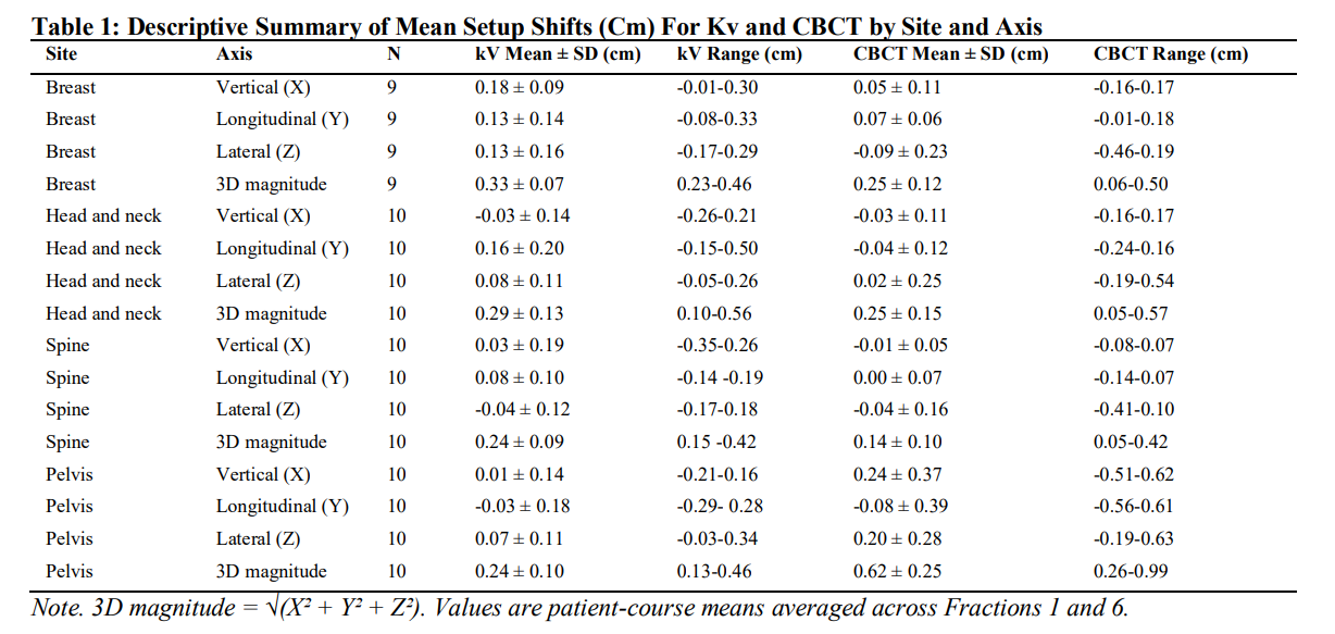
Cone-beam computed tomography (CBCT) is widely used in image-guided radiotherapy (IGRT) because it provides volumetric verification of patient setup, but routine use may be difficult to sustain in resource-constrained settings. Planar kilovoltage (kV) imaging is faster and simpler, although its positioning accuracy may vary across treatment sites. The research aimed to assess the impact of kV imaging on patient positioning accuracy across different treatment sites compared with CBCT. This prospective within-patient method-comparison study included 39 patient-courses treated at the University of Nigeria Teaching Hospital, comprising breast (n = 9), head and neck (n = 10), spine (n = 10), and pelvis (n = 10).Paired CBCT and orthogonal kV images were acquired on Fractions 1 and 6.Translational couch shifts in the vertical, longitudinal, and lateral directions were analysed using paired t-tests, Bland-Altman analysis, and clinical agreement rates within ±0.3 cm.Interobserver reliability of kV matching was assessed using intraclass correlation coefficient. Mean three-dimensional differences between kV and CBCT were not significant for breast (0.07 cm; p = 0.158), head and neck (0.04 cm; p = 0.584), and spine (0.095 cm; p= 0.099), but were significant for pelvis (-0.38 cm; p = 0.002).Agreement within ±0.3 cm was highest in spine (100%), followed by head and neck (80-100%) and breast (66.7–88.9%), while pelvis showed the lowest agreement (50-70%). kV interobserver reliability was good to excellent (ICC = 0.724-0.945). kV imaging can provide clinically acceptable positioning accuracy in relatively rigid treatment sites, especially spine and head and neck, but pelvic cases still require CBCT support.References
Çini, N., & Biçakçi, B. C. (2023). Evaluation of set-up shift values detected in CBCT and kV images taken on set-up day and interfraction treatment days in patients with laryngeal cancer who underwent image-guided radiotherapy. Demiroglu Science University Florence Nightingale Journal of Medicine, 9(1), 12-18. https://doi.org/10.54614/DSFNJM.2023.47381
Cai, J., Peng, X., & Lu, J. (2025). Dosimetric and radiobiological impact of patient setup errors in intensity-modulated radiotherapy for oesophageal cancer. Technology in Cancer Research & Treatment, 24, Article 15330338241307960. https://doi.org/10.1177/15330338241307960
Ding, G. X., Alaei, P., Curran, B., Flynn, R., Gossman, M., Mackie, T., Miften, M., Morin, R., Xu, X. G., & Zhu, T. (2018). Image guidance doses delivered during radiotherapy: Quantification, management, and reduction. Medical Physics, 45(5), e84-e99. https://doi.org/10.1002/mp.12824
Elakshar, J., Tsui, M., Kucharczyk, M., Tomic, N., Fawaz, Z., Bahoric, B., Papayanatos, J., Chaddad, A., & Niazi, T. (2019). Does interfraction cone beam computed tomography improve target localization in prostate bed radiotherapy? Technology in Cancer Research & Treatment, 18, Article 1533033819829704. https://doi.org/10.1177/1533033819829704
Eren, M. F., Çolpan Öksüz, D., Sayan, M., Karaçam, S., Vergalasova, I., Ay Eren, A., & Öner Dinçbaş, F. (2020). Comparison of kV orthogonal radiographs and kV cone beam computed tomography image-guided radiotherapy methods with and without implanted fiducials in prostate cancer. Cureus, 12(8), e9916. https://doi.org/10.7759/cureus.9916
Federal Ministry of Health. (2022). National cancer control programme report 2018-2022. Federal Ministry of Health.
Grégoire, V., Guckenberger, M., Haustermans, K., Lagendijk, J. J. W., Ménard, C., Pötter, R., Slotman, B. J., Tanderup, K., Thorwarth, D., van Herk, M., & Zips, D. (2020). Image guidance in radiation therapy for better cure of cancer. Molecular Oncology, 14(7), 1470-1491. https://doi.org/10.1002/1878-0261.12751
HopeJohnson, T., Parkes, J., Prajogi, G., Sullivan, R., Vanderpuye, V., & Aggarwal, A. (2025). Strengthening capacity in radiotherapy skills to deliver high-quality treatments in low- and middle-income countries. International Journal of Radiation Oncology, Biology, Physics, 121(4), 856-862. https://doi.org/10.1016/j.ijrobp.2024.10.034
Iliopoulos, P., Simopoulou, F., Simopoulos, V., Kyrgias, G., & Theodorou, K. (2023). Review on cone beam computed tomography (CBCT) dose in patients undergoing image-guided radiotherapy (IGRT). In Advances in dosimetry and new trends in radiopharmaceuticals. IntechOpen. https://doi.org/10.5772/intechopen.111086
Jaffray, D. A. (2012). Image-guided radiotherapy: From current concept to future perspective. Nature Reviews Clinical Oncology, 9(12), 688-699. https://doi.org/10.1038/nrclinonc.2012.194
Kang, H., Lovelock, D. M., & Yorke, E. D. (2010). Accurate positioning for head and neck cancer patients using 2D and 3D image guidance. Journal of Applied Clinical Medical Physics, 11(4), Article 3270. https://doi.org/10.1120/jacmp.v11i4.3270
Koo, T. K., & Li, M. Y. (2016). A guideline of selecting and reporting intraclass correlation coefficients for reliability research. Journal of Chiropractic Medicine, 15(2), 155-163. https://doi.org/10.1016/j.jcm.2016.02.012
Kuo, Y., Liang, J., Chen, G., & Chien, C. (2021). Safety of image-guided radiotherapy in definitive radiotherapy for localised prostate cancer: A population-based analysis. The British Journal of Radiology, 96(1121), 20200870. https://doi.org/10.1259/bjr.20200870
Quinn, L., Tryposkiadis, K., Deeks, J., et al. (2023). Interobserver variability studies in diagnostic imaging: A methodological systematic review. The British Journal of Radiology, 96(1148), 20220972. https://doi.org/10.1259/bjr.20220972
Sarma, G., Kashyap, H., Medhi, P. P., Kalita, R., & Lahkar, D. (2024). Unravelling the landscape of image-guided radiotherapy: A comprehensive overview. Palliative Medicine in Practice, 18(4), 228-233. https://doi.org/10.5603/PMP.101197
Topolnjak, R., Bijhold, J., Sonke, J. J., & Remeijer, P. (2010). Patient setup verification using cone-beam CT for breast radiotherapy. International Journal of Radiation Oncology, Biology, Physics, 77(5), 1521-1529. https://doi.org/10.1016/j.ijrobp.2009.10.029
van Herk, M., Remeijer, P., Rasch, C., & Lebesque, J. V. (2000). The probability of correct target dosage: Dose-population histograms for deriving treatment margins in radiotherapy. International Journal of Radiation Oncology, Biology, Physics, 47(4), 1121-1135. https://doi.org/10.1016/S0360-3016(00)00518-6
Wang, K., & Tepper, J. E. (2021). Radiation therapy-associated toxicity: Etiology, management and prevention. CA: A Cancer Journal for Clinicians, 71(5), 437-454. https://doi.org/10.3322/caac.21689
Wang, W., Yu, T., Xu, M., Shao, Q., Zhang, Y., & Li, J. (2019). Setup error assessment and correction in planar kV image versus cone beam CT image-guided radiation therapy: A clinical study of early breast cancer treated with external beam partial breast irradiation. Technology in Cancer Research & Treatment, 18, Article 1533033819853847. https://doi.org/10.1177/1533033819853847
Webster, A., Appelt, A. L., & Eminowicz, G. (2020). Image-guided radiotherapy for pelvic cancers: A review of current evidence and clinical utilisation. Clinical Oncology, 32(12), 805-816. https://doi.org/10.1016/j.clon.2020.08.002
Wilson, L., Hadjipanteli, A., Ostergaard, D. E., Bogaert, E., Brown, C. F., DeJong, R., Earley, J., Edouard, M., Khan, M., Lindsay, J., Van Der Himst, J., Ding, G. X., Wood, T., Aznar, M. C., Jornet, N., & Ntentas, G. (2025). Cone beam CT dose optimisation: A review and expert consensus by the 2022 ESTRO Physics Workshop IGRT working group. Radiotherapy and Oncology, 208, 1-9. https://doi.org/10.1016/j.radonc.2025.110174
Downloads
Published
Issue
Section
Categories
License
Copyright (c) 2026 Abdullahi Shuaibu, Charles Ugwoke Eze, Anakwue Angel-Mary, Mohammed Yusuf Mohammed, Abdurrahman Umaru, Everistus Obinna Abonyi, Inwang Edet Usoro

This work is licensed under a Creative Commons Attribution 4.0 International License.

