
Biosynthesis of Calcium Oxide Nanoparticles Using Jatropha tanjorensis Leaf Extract and the Evaluation of its Antibacterial Efficacy Against Multidrug Resistance Uropathogens and Haemocompatibility
DOI:
https://doi.org/10.33003/fjs-2026-1017-6022Keywords:
Green Synthesis, Calcium Oxide Nanoparticles, Jatropha tanjorensis, Antibacterial Activity, Uropathogens, HaemocompatibilityAbstract
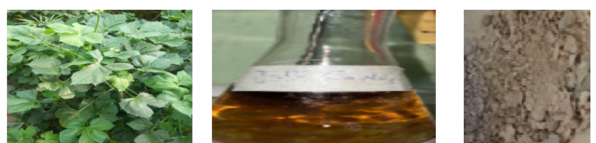
The biosynthesis of metal oxide nanoparticles using phytochemicals from plant extracts has been recognized as a sustainable, low-toxicity alternative to chemically synthesized antimicrobials. It provides an environmentally friendly approach to combat the growing burden of multidrug-resistant (MDR) pathogens. This study investigates the biosynthesis of calcium oxide nanoparticles (CaONPs) using aqueous leaf extract of Jatropha tanjorensis, an indigenous Nigerian medicinal plant rich in phytochemicals capable of chelating and reducing Ca²⁺ ions, and evaluates their antibacterial and haemocompatibility profiles against pathogens isolated from urine samples. CaONPs were synthesized by combining CaCl₂ solution with the leaf extract under alkaline conditions, followed by centrifugation and calcination at 700 °C. The nanoparticles were characterized using ultraviolet-visible spectrophotometry (UV-Vis), Fourier transform infrared (FTIR), X-ray diffraction (XRD) and scanning electron microscopy (SEM). Antibacterial activity was assessed against four uropathogenic isolates, Klebsiella pneumoniae, Proteus mirabilis, Bacillus cereus and Staphylococcus aureus, using agar diffusion and broth microdilution to determine MIC and MBC. Biocompatibility was evaluated using a haemolytic assay on human erythrocytes. Characterization confirmed nanoparticle formation, phytochemical capping, heterogeneous surface morphology and particle dimensions within the nanoparticle size range. All isolates showed concentration-dependent bactericidal activity, with MIC and MBC values ranging from 1.88–3.75 µg/mL for P. mirabilis and 7.50–15.00 µg/mL for the remaining isolates, with inhibition zones of 15.20–22.00 mm. Haemolytic assays showed a favorable safety margin, with an estimated LC₅₀ of 436.973 µg/mL, indicating minimal haemolytic activity within the effective antibacterial range. These findings position J. tanjorensis-mediated CaONPs as promising, biocompatible option for topical or catheter-coating antimicrobial applications.
References
Aghayan, N. S., Seyfi, J., Asadollahzadeh, M. J., Davachi, S. M., & Hasani, M. (2022). Preparation and characterization of antibacterial chitosan nanocomposites loaded with cellulose/chitosan nanofibers and essential oils. Journal of Thermoplastic Composite Materials. https://doi.org/10.1177/09673911221098732
Algadi, H., Alhoot, M. A., & Yaaqoob, L. A. (2025). Systematic review of antibacterial potential in calcium oxide and silicon oxide nanoparticles for clinical and environmental infection control. Journal of Applied Biomedicine, 23(1), 1–11. https://doi.org/10.32725/jab.2025.001
Azam, A., Ahmed, A. S., Oves, M., Khan, M. S., Habib, S. S., & Memic, A. (2012). Antimicrobial activity of metal oxide nanoparticles against Gram-positive and Gram-negative bacteria: A comparative study. International Journal of Nanomedicine, 7, 6003–6009. https://doi.org/10.2147/IJN.S35347
Böhmert, L., Niemann, B., Lichtenstein, D., Juling, S., Lampen, A., & Laux, P. (2019). The hemocompatibility of nanoparticles: A review of cell–nanoparticle interactions and hemostasis. Cells, 8(10), 1209. https://doi.org/10.3390/cells8101209
Cheesbrough, M. (2006). District laboratory practice in tropical countries (Part 2, 2nd ed.). Cambridge University Press.
Clinical and Laboratory Standards Institute (2018). Methods for Dilution Antimicrobial Susceptibility Tests for Bacteria That Grow Aerobically; Approved Standard 11th Edition. Wayne, PA: CLSI.
Dobrovolskaia, M. A., Clogston, J. D., Neun, B. W., Hall, J. B., Patri, A. K., & McNeil, S. E. (2008). Method for analysis of nanoparticle hemolytic properties in vitro. Nano Letters, 8(8), 2180–2187. https://doi.org/10.1021/nl080511k
El-Seedi, H. R., Omara, M. S., Omar, A. H., Elakshar, M. M., Shoukhba, Y. M., Duman, H., Karav, S., Rashwan, A. K., El-Seedi, A. H., Altaleb, H. A., Gao, H., Saeed, A., Jefri, O. A., Guo, Z., & Khalifa, S. A. M. (2024). Updated review of metal nanoparticles fabricated by green chemistry using natural extracts: Biosynthesis, mechanisms, and applications. Bioengineering, 11(11), Article 1095. https://doi.org/10.3390/bioengineering11111095
Falodun, A., Udu-Cosi, A. A., Erharuyi, O., Imieje, V., Falodun, J. E., Agbonlahor, O., & Hamann, M. T. (2014). Jatropha tanjorensis—Review of phytochemistry, pharmacology, and pharmacotherapy. Journal of Pharmaceutical and Allied Sciences, 10(3). https://doi.org/10.4314/jophas.v10i3
Futoma-Kołoch, B., Sarowska, J., Abd El-Salam, M., Miñana-Galbis, D., Drabová, B., Guz-Regner, K., Wiśniewska, P., & Kryniewska, V. (2025). Current insights into antibiotic resistance in uropathogenic Escherichia coli and interventions using selected bioactive phytochemicals. Antibiotics, 14(12), Article 1242. https://doi.org/10.3390/antibiotics14121242
Gedda, G., Pandey, S., Lin, Y.-C., & Wu, H.-F. (2015). Antibacterial effect of calcium oxide nano-plates fabricated from shrimp shells. Green Chemistry, 17(6), 3276–3280. https://doi.org/10.1039/C5GC00615E
Harborne, J. B. (1998). Phytochemical methods: A guide to modern techniques of plant analysis (3rd ed.). Chapman & Hall.
Hunter, J. D. (2007). Matplotlib: A 2D graphics environment. Computing in Science & Engineering, 9(3), 90–95. https://doi.org/10.1109/MCSE.2007.55
Jadhav, V., Bhagare, A., Wahab, S., Lokhande, D., Vaidya, C., Dhayagude, A., Khalid, M., Aher, J., Mezni, A., & Dutta, M. (2022). Green synthesized calcium oxide nanoparticles using leaves aqueous extract of Moringa oleifera and evaluation of their antibacterial activities. Journal of Nanomaterials, 2022, Article 9047507. https://doi.org/10.1155/2022/9047507
Khalid, R., Din, M.I., Parveen, A. & Hussain, Z. (2025). A Critical Review on Green Synthesis of Calcium Oxide Nanoparticles and Calcium Based Nanocomposites. BioNanoSci. 15(390). https://doi.org/10.1007/s12668-025-02013-2
Khan, A. U., Hussain, T., Abdullah, Khan, M. A., Almostafa, M. M., Younis, N. S., & Yahya, G. (2023). Antibacterial and antibiofilm activity of Ficus carica-mediated calcium oxide (CaONPs) phyto-nanoparticles. Molecules, 28(14), Article 5553. https://doi.org/10.3390/molecules28145553
Kumari, M., Sarkar, B., & Mukherjee, K. (2023). Nanoscale calcium oxide and its biomedical applications: A comprehensive review. Biocatalysis and Agricultural Biotechnology, 47, Article 102506. https://doi.org/10.1016/j.bcab.2022.102506
Lawrie, G., Keen, I., Drew, B., Chandler-Temple, A., Rintoul, L., Fredericks, P., & Grøndahl, L. (2007). Interactions between alginate and chitosan biopolymers characterized using FTIR and XPS. Biomacromolecules, 8(8), 2533–2541. https://doi.org/10.1021/bm070014y
Lithi, I. J., Nakib, K. I. A., Chowdhury, A. M. S., & Hossain, M. S. (2025). A review on the green synthesis of metal (Ag, Cu, and Au) and metal oxide (ZnO, MgO, Co₃O₄, and TiO₂) nanoparticles using plant extracts for developing antimicrobial properties. Nanoscale Advances, 7(9), 2446–2473. https://doi.org/10.1039/D5NA00037H
Malehmir, S., Esmaili, M. A., Khaksary Mahabady, M., Sobhani-Nasab, A., Atapour, A., Ganjali, M. R., Ghasemi, A., & Moradi Hasan-Abad, A. (2023). A review: Hemocompatibility of magnetic nanoparticles and their regenerative medicine, cancer therapy, drug delivery, and bioimaging applications. Frontiers in Chemistry, 11, Article 1249134. https://doi.org/10.3389/fchem.2023.1249134
Mazher, M., Ishtiaq, M., Hamid, B., Haq, S. M., Mazhar, A., Bashir, F., Mazhar, M., Mahmoud, E. A., Casini, R., Alataway, A., Dewidar, A. Z., & Elansary, H. O. (2023). Biosynthesis and characterization of calcium oxide nanoparticles from Citrullus colocynthis fruit extracts: Their biocompatibility and bioactivities. Materials, 16(7), Article 2768. https://doi.org/10.3390/ma16072768
Mito, D., Okihara, S.-i., Kurita, M., Hatayama, N., Yoshino, Y., Watanabe, Y., & Ishii, K. (2024). Leveraging broad-spectrum fluorescence data and machine learning for high-accuracy bacterial species identification. Journal of Biophotonics, 17(12), e202400300. https://doi.org/10.1002/jbio.202400300
Musa, H., & Abubakar, A. A. (2023). Synthesis, Characterization and Application of Silver-nanoparticles-Embedded Alkyd Resins Derived from Jathropha Seed Oil. FUDMA Journal of Sciences, 3(1), 312-320
Naik, T. S. S. K., Singh, S., Narasimhappa, P., Varshney, R., Singh, J., Khan, N. A., Zahmatkesh, S., Ramamurthy, P. C., Shehata, N., Kiran, G. N., & Sunil, K. (2023). Green and sustainable synthesis of CaO nanoparticles: Its solicitation as a sensor material and electrochemical detection of urea. Scientific Reports, 13, Article 19948. https://doi.org/10.1038/s41598-023-46728-2
Neun, B. W., Ilinskaya, A. N., & Dobrovolskaia, M. A. (2018). Updated method for in vitro analysis of nanoparticle hemolytic properties. In Characterization of Nanoparticles Intended for Drug Delivery Methods in Molecular Biology, Vol. 1682, pp. 91–102. Springer. https://doi.org/10.1007/978-1-4939-7352-1_9
Nguyen, N. T. T., Nguyen, L. M., Nguyen, T. T. T., Nguyen, T. T., Nguyen, D. T. C., & Tran, T. V. (2022). Formation, antimicrobial activity, and biomedical performance of plant-based nanoparticles: A review. Environmental Chemistry Letters, 20(4), 2531–2571. https://doi.org/10.1007/s10311-022-01425-w
Oghenefejiro, O. P., Ikhuoria, E. U., Ehigie, A. C., Osarobo, J. A., & Ifijen, I. H. (2025). Green synthesis of calcium oxide nanoparticles using mango (Mangifera indica) leaf extract: A sustainable approach for enhanced antimicrobial applications. Biomedical Materials & Devices, 4, 3533–3544. https://doi.org/10.1007/s44174-025-00375-5
Pankey, G. A., & Sabath, L. D. (2004). Clinical relevance of bacteriostatic versus bactericidal mechanisms of action in the treatment of Gram-positive bacterial infections. Clinical Infectious Diseases, 38(6), 864–870. https://doi.org/10.1086/381972
Patterson, A. L. (1939). The Scherrer formula for X-ray particle size determination. Physical Review, 56(10), 978–982.
Rana, A., Pathak, S., Lim, D.-K., Kim, S.-K., Srivastava, R., Sharma, S. N., & Verma, R. (2023). Recent advancements in plant- and microbe-mediated synthesis of metal and metal oxide nanomaterials and their emerging antimicrobial applications. ACS Applied Nano Materials, 6(10), 8106–8134. https://doi.org/10.1021/acsanm.3c01351
Roy, B., Das, T., & Bhattacharyya, S. (2023). Overview on old and new biochemical test for bacterial identification. Journal of Surgical Case Reports and Images, 6(5), 1–11.
Shandhiya, M., Janarthanan, B., & Sharmila, S. (2024). A comprehensive review on antibacterial analysis of natural extract-based metal and metal oxide nanoparticles. Archives of Microbiology, 206, Article 52. https://doi.org/10.1007/s00203-023-03743-1
Wiegand, I., Hilpert, K., & Hancock, R. E. W. (2008). Agar and broth dilution methods to determine the minimal inhibitory concentration (MIC) of antimicrobial substances. Nature Protocols, 3(2), 163–175. https://doi.org/10.1038/nprot.2007.521
Yoonus, J., Resmi, R., & Beena, B. (2021). Greener nanoscience: Piper betel leaf extract-mediated synthesis of CaO nanoparticles and evaluation of its antibacterial and anticancer activity. Materials Today: Proceedings, 41, 535–540. https://doi.org/10.1016/j.matpr.2020.05.246
Yusuf, C. S., Bwatanglang, I. B., Tizhe, T. D., Tosin, P. E., & Felister, M. R. (2021). Ethnobotany and phytochemical analysis of Jatropha tanjorensis in Mubi, Adamawa State, Nigeria. International Journal of Frontiers in Life Science Research, 1(1), 40–47. https://doi.org/10.53294/ijflsr.2021.1.1.0035
Downloads
Published
Issue
Section
Categories
License
Copyright (c) 2026 Mirabel N. Ekeogu, Sandra N. Iweriolor, Bright O. Uba

This work is licensed under a Creative Commons Attribution 4.0 International License.