Embedding Media in Histopathology: A Narrative Review of Paraffin Wax, and Natural Alternatives
DOI:
https://doi.org/10.33003/fjs-2026-1015-5972Keywords:
Histopathology, Tissue embedding, Paraffin wax, Natural waxes, Sustainable laboratory practicesAbstract
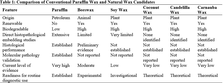
Tissue embedding is a critical step in histopathological processing because it provides the structural support required for thin sectioning, staining, and microscopic examination. For more than a century, paraffin wax has remained the standard embedding medium because of its excellent tissue infiltration, consistent ribbon formation, preservation of tissue morphology, long-term storage stability, and compatibility with routine histology, immunohistochemistry, and molecular diagnostic techniques. However, concerns regarding its petroleum-derived origin, environmental impact, and sustainability have stimulated interest in renewable natural waxes as alternatives. This narrative review critically evaluated paraffin wax and selected natural embedding media, including beeswax, soy wax, coconut wax, candelilla wax, carnauba wax, rice bran wax, palm wax, and bayberry wax. Relevant peer-reviewed literatures were synthesized to compare their physicochemical properties, embedding performance, advantages, limitations, and suitability for routine histopathological practice. Evidence showed that paraffin wax continues to provide superior overall performance and remains the benchmark for diagnostic tissue embedding. Among the natural alternatives, beeswax demonstrated the strongest direct evidence for tissue embedding, while soy wax showed potential in modified formulations. Evidence supporting coconut, candelilla, carnauba, rice bran, palm, and bayberry waxes remains largely indirect and based on physicochemical or industrial studies rather than validated histopathological applications. Key challenges include limited comparative studies, lack of standardized evaluation protocols, variability in natural wax composition, and inadequate validation with ancillary diagnostic techniques. Overall, natural waxes offer promising sustainable alternatives, but current evidence is insufficient to support their routine replacement of paraffin wax in diagnostic histopathology, highlighting the need for standardized experimental studies.
References
(2021). Standard techniques for formalin-fixed paraffin-embedded tissue: A pathologist’s perspective. Iraqi Journal of Veterinary Sciences, 35(Supplement I-III), 127–135. https://doi.org/10.33899/ijvs.2021.131918.2023
Aranda-Ledesma, N. E., Bautista-Hernández, I., Rojas, R., Aguilar-Zárate, P., Medina-Herrera, N. P., Castro-López, C., & Guadalupe Martínez-Ávila, G. C. (2022). Candelilla wax: Prospective suitable applications within the food field. LWT, 159, 113170. https://doi.org/10.1016/j.lwt.2022.113170
Atreya, M., Marinick, G., Baumbauer, C., Dikshit, K. V., Liu, S., Bellerjeau, C., Nielson, J., Khorchidian, S., Palmgren, A., Sui, Y., Bardgett, R., Baumbauer, D., Bruns, C. J., Neff, J. C., Arias, A. C., & Whiting, G. L. (2022). Wax blends as tunable encapsulants for soil-degradable electronics. ACS Applied Electronic Materials, 4(10), 4912–4920. https://doi.org/10.1021/acsaelm.2c00833
Bai, Z., Zhang, D., Gao, Y., Tao, B., Zhang, D., Bao, S., Enninful, A., Wang, Y., Li, H., Su, G., Tian, X., Zhang, N., Xiao, Y., Liu, Y., Gerstein, M., Li, M., Xing, Y., Lu, J., Xu, M. L., & Fan, R. (2024). Spatially exploring RNA biology in archival formalin-fixed paraffin-embedded tissues. Cell, 187(23), 6760–6779.e24. https://doi.org/10.1016/j.cell.2024.09.001
Bass, B. P., Engel, K. B., Greytak, S. R., & Moore, H. M. (2014). A review of preanalytical factors affecting molecular, protein, and morphological analysis of formalin-fixed, paraffin-embedded (FFPE) tissue: How well do you know your FFPE specimen? Archives of Pathology & Laboratory Medicine, 138(11), 1520–1530. https://doi.org/10.5858/arpa.2013-0691-ra
Bucio, A., Moreno-Tovar, R., Bucio, L., Espinosa-Dávila, J., & Anguebes-Franceschi, F. (2021). Characterization of beeswax, candelilla wax and paraffin wax for coating cheeses. Coatings, 11(3), 261. https://doi.org/10.3390/coatings11030261
Copper, J. E., Budgeon, L. R., Foutz, C. A., van Rossum, D. B., Vanselow, D. J., Hubley, M. J., Clark, D. P., Mandrell, D. T., & Cheng, K. C. (2018). Comparative analysis of fixation and embedding techniques for optimized histological preparation of zebrafish. Comparative Biochemistry and Physiology Part C: Toxicology & Pharmacology, 208, 38–46. https://doi.org/10.1016/j.cbpc.2017.11.003
Dewi, H., Quzwain, F., & Wulansari, N. (2022). Histology slide quality comparative study; impregnation and embedding using beeswax and paraffin. Jambi Medical Journal : Jurnal Kedokteran Dan Kesehatan, 10(2), 291–298. https://online-journal.unja.ac.id/kedokteran/article/view/19242
Gupta, N., Chandra, S., Gupta, S., Samadi, F., & Chauhan, M. (2023). Comparative diagnostic accuracy of paraffin wax versus beeswax for tissue embedding. International Journal of Medical Toxicology & Legal Medicine, 26(1and2), 105–109. https://doi.org/10.5958/0974-4614.2023.00019.0
Hasegawa, Y., Mark Welch, J. L., Rossetti, B. J., & Borisy, G. G. (2017). Preservation of three-dimensional spatial structure in the gut microbiome. PLOS ONE, 12(11), e0188257. https://doi.org/10.1371/journal.pone.0188257
Isaac, U. E., Oyo-Ita, E., Igwe, N. P., & Ije, E. L. (2023). Preparation of histology slides and photomicrographs: Indispensable techniques in anatomic education. Anatomy Journal of Africa, 12(1), 2252–2262. https://doi.org/10.4314/aja.v12i1.1
İnan, Z. D. Ş., Hamutoğlu, R., & Saraydın, S. Ü. (2025). Comparative analysis of paraffin and JB-4 embedding techniques in light microscopy. Biology Methods and Protocols, 10(1). https://doi.org/10.1093/biomethods/bpaf071
Kharomah, I. F., & Nailufar, Y. (2024). Exploring coconut oil as a green substitute for xylene in histology clearing. Journal of Public Health Sciences, 3(01), 1–9. https://doi.org/10.56741/jphs.v3i01.446
Lemaire, R., Desmons, A., Tabet, J. C., Day, R., Salzet, M., & Fournier, I. (2007). Direct analysis and MALDI imaging of formalin-fixed, paraffin-embedded tissue sections. Journal of Proteome Research, 6(4), 1295–1305. https://doi.org/10.1021/pr060549i
Likhithaswamy, H. R., Madhushankari, G. S., Selvamani, M., Mohan Kumar, K. P., Kokila, G., & Mahalakshmi, S. (2022). Assessing the quality of long-term stored tissues in formalin and in paraffin-embedded blocks for histopathological analysis. Journal of Microscopy and Ultrastructure, 10(1), 23–29. https://doi.org/10.4103/jmau.jmau_53_20
Marinho, P. F., & Hanscheid, T. (2023). A simple heat-based alternative method for deparaffinization of histological sections significantly improves acid-fast staining results for Mycobacteria in tissue. MethodsX, 10, 102079. https://doi.org/10.1016/j.mex.2023.102079
Mazzoni, T. S., & Quagio‐Grassiotto, I. (2021). In totum deparaffinization of biological samples and re‐embedded in historesin for better diagnostic. Microscopy Research and Technique, 84(7), 1362–1367. https://doi.org/10.1002/jemt.23695
Nilcham, P., Afify, M., Schaaps, N., Neu, C., Shahin, R., Prescher, A., Vogt, F. J., & Turoni-Glitz, A. (2025). Histo-LOOP: A novel embedding tool for standardizing, simplifying, and advancing histological tissue preparation. Journal of Histochemistry & Cytochemistry, 73(3–4), 97–107. https://doi.org/10.1369/00221554251329978
Núñez-García, I. C., Rodríguez-Flores, L. G., Guadiana-De-Dios, M. H., González-Hernández, M. D., Martínez-Ávila, G. C. G., Gallegos-Infante, J. A., González-Laredo, R., Rosas-Flores, W., Martínez-Gómez, V. J., Rojas, R., Villanueva-Fierro, I., & Rutiaga-Quiñones, M. (2022). Candelilla wax extracted by traditional method and an ecofriendly process: Assessment of its chemical, structural and thermal properties. Molecules, 27(12), 3735. https://doi.org/10.3390/molecules27123735
Ridolfi, M., Paudice, M., Salvi, S., Valle, L., Gualco, M., Perasole, A., Anselmi, L., Fiocca, R., Mastracci, L., & Grillo, F. (2019). Agar pre-embedding of small skin biopsies: Real-life benefits and challenges in high throughput pathology laboratories. Journal of Clinical Pathology, 72(6), 448–451. https://doi.org/10.1136/jclinpath-2018-205680
Romero, S., Minari, R. J., & Collins, S. E. (2021). Bio-paraffin from soybean oil as eco-friendly alternative to mineral waxes. Industrial & Engineering Chemistry Research, 60(15), 5364–5373. https://doi.org/10.1021/acs.iecr.1c00322
Shimizu, T., Tanabe, T., Kachi, H., & Shibata, M. (2022). Enhancement of the gel hardness of candelilla wax through the addition of long-chain Ester Wax Behenyl Behenate. Journal of Oleo Science, 71(12), 1725–1733. https://doi.org/10.5650/jos.ess22175
Taffon, C., Naciu, A. M., Bonfiglio, R., Palumbo, V., Maricchiolo, G., Morano, V., Salducci, M., Stigliano, S., Palermo, A., Di Matteo, F. M., & Crescenzi, A. (2024). From sampling to cellblock: The fully automated journey of cytological specimens. Diagnostic Cytopathology, 52(11), 611–616. https://doi.org/10.1002/dc.25366
Taqi, S. A., Sami, S. A., Sami, L. B., & Zaki, S. A. (2018). A review of artifacts in histopathology. Journal of Oral and Maxillofacial Pathology, 22(2), 279. https://doi.org/10.4103/jomfp.jomfp_125_15
Thajudeen, A., Srinivasan, S., Govindarajan, G., & Shanmugam, A. (2022). A comparative study of efficacy of coconut oil, lemon water and dishwashing liquid as surrogates to xylene. Environmental Analysis Health and Toxicology, 37(3), e2022026. https://doi.org/10.5620/eaht.2022026
Thamilselvan, S., Sherlin, H. J., Jayaraj, G., Don, K. R., & Santhanam, A. (2021). Cedarwood oil as an alternative to xylene as a clearing agent in histopathological tissue processing – a comparative study. Journal of Oral and Maxillofacial Pathology, 25(2), 299–305. https://doi.org/10.4103/0973-029x.325232
van der Lem, T., de Bakker, M., Keuck, G., & Richardson, M. K. (2021). Wilhelm His Sr. and the development of paraffin embedding. Der Pathologe, 42(S1), 55–61. https://doi.org/10.1007/s00292-021-00947-4
Varshini, M., Pushparaj, S., & Mangesh, S. (2026). Advancing green chemistry in histopathology: A comparative study of coconut oil and turpentine oil as deparaffinizing agents. Indian Journal of Pathology and Microbiology, 69(1), 101–105. https://doi.org/10.4103/ijpm.ijpm_836_24
Vini, J., & Antony, M. (2017). Impregnation and embedding using bees wax and paraffin wax in oral tissue samples: A comparative study. International Journal of Orofacial Biology, 1(13–5). https://doi.org/10.4103/ijofb.ijofb_3_17
Wang, Q., Gu, R., Olm, F., Bèchet, N. B., & Lindstedt, S. (2024). Xylene versus isopropanol for paraffin wax processing of lung tissue. Applied Sciences, 14(5), 1726. https://doi.org/10.3390/app14051726
Zhanmu, O., Yang, X., Gong, H., & Li, X. (2020). Paraffin-embedding for large volume bio-tissue. Scientific Reports, 10(1), 12639. https://doi.org/10.1038/s41598-020-68876-5
Downloads
Published
Issue
Section
Categories
License
Copyright (c) 2026 Gabriel A. Funsho-Agun, Ifeyinwa Nokwai-Ikeji

This work is licensed under a Creative Commons Attribution 4.0 International License.