
Indications and Spectrum of Radiological Findings on Intravenous Urography in a Resource-Limited Tertiary Hospital in Northwest Nigeria: A six-Year Retrospective Review
DOI:
https://doi.org/10.33003/fjs-2026-1010-5245Keywords:
Intravenous Urography, Radiological Findings, Hydronephrosis, Urolithiasis, Retrospective Study, NigeriaAbstract
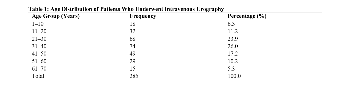
Intravenous urography (IVU) remains an important imaging modality in many resource-limited healthcare settings despite the increasing availability of computed tomography urography and other advanced cross-sectional imaging techniques. This retrospective analysis evaluated the indications for IVU and the spectrum of radiological findings among patients who underwent the examination in a tertiary healthcare facility in Northwest Nigeria. A hospital-based retrospective descriptive analysis was conducted at the Department of Radiology, Federal Medical Centre Birnin Kebbi, now Federal Teaching Hospital Birnin Kebbi, Nigeria. Records of 285 patients who underwent IVU between January 2019 and December 2024 were reviewed. Demographic characteristics, clinical indications, and radiological findings were extracted from archived radiology records and analysed using SPSS version 22. Ages ranged from 1 to 70 years, with a mean age of 35.8 ± 14.6 years. Males accounted for 204 (71.6%) patients. The commonest indications were suspected urinary tract obstruction (33.7%), suspected urinary calculi (24.9%), and haematuria (15.4%). Hydronephrosis (27.4%), urinary calculi (22.5%), and hydroureter (13.7%) were the most frequent findings. Significant associations were observed between sex and urinary calculi (χ² = 4.43, p = 0.035), clinical indication and hydronephrosis (χ² = 33.01, p < 0.001), clinical indication and urinary calculi (χ² = 25.81, p < 0.001), and mean age and urinary calculi (t = 2.31, p = 0.022). IVU remains valuable in resource-limited settings, particularly for obstructive uropathy and urolithiasis.
References
Abou-El-Ghar, M., Refaie, H., Sharaf, D., & El-Diasty, T. (2014). Diagnosing urinary tract abnormalities: Intravenous urography or CT urography? Reports in Medical Imaging, 7, 55–63. https://doi.org/10.2147/RMI.S35592
Akagbue, V. N., & Wekhe, C. (2023). Intravenous urography findings among patients presenting to Rivers State University Teaching Hospital: A two-year retrospective study. International Journal of Medical Science and Clinical Research Studies, 3(11), 2858–2864. https://doi.org/10.47191/ijmscrs/v3-i11-60
Aklan, H. M., & Mikhlafy, A. (2018). Analysis of intravenous urography findings in a tertiary reference center. The Eurasian Journal of Medicine, 50(2), 71–74. https://doi.org/10.5152/eurasianjmed.2018.170304
Chen, M. Y. M., Zagoria, R. J., & Dyer, R. B. (1997). Radiologic findings in acute urinary tract obstruction. The Journal of Emergency Medicine, 15(3), 339–343. https://doi.org/10.1016/S0736-4679(97)82536-X
Cowan, N. C. (2012). CT urography for hematuria. Nature Reviews Urology, 9(4), 218–226. https://doi.org/10.1038/nrurol.2012.32
Dyer, R. B., Chen, M. Y. M., & Zagoria, R. J. (2001). Intravenous urography: Technique and interpretation. RadioGraphics, 21(4), 799–824. https://doi.org/10.1148/radiographics.21.4.g01jl26799
Kawashima, A., Vrtiska, T. J., LeRoy, A. J., Hartman, R. P., McCollough, C. H., & King, B. F. (2004). CT urography. RadioGraphics, 24(Suppl. 1), S35–S54. https://doi.org/10.1148/rg.24si045513
Kawooya, M. G. (2012). Training for rural radiology and imaging in Sub-Saharan Africa: Addressing the mismatch between services and population. Journal of Clinical Imaging Science, 2, 37. https://doi.org/10.4103/2156-7514.97747
Muhammad, Y. U., Sada, A. N., & Mukhtar, U. F. (2026). Enhancing diagnostic accuracy in Nigerian hospitals: Analysis of deep learning applications in medical imaging. FUDMA Journal of Sciences, 10(ANB-K), 134–141. https://doi.org/10.33003/fjs-2026-10(ANB-K)-5193
National Population Commission. (2010). 2006 population and housing census of the Federal Republic of Nigeria: Priority tables, volume III. National Population Commission.
Ogunbiyi, O. A., & Nzeh, D. (1984). Intravenous urography in paediatric and adolescent practice. Nigerian Journal of Paediatrics, 11(4), 107–114.
Pearle, M. S., Calhoun, E. A., & Curhan, G. C. (2005). Urologic diseases in America project: Urolithiasis. The Journal of Urology, 173(3), 848–857. https://doi.org/10.1097/01.ju.0000152082.14384.d7
Raza, M., Khan, S. F., Ahmed, S., & Zameer, S. (2011). Diagnostic yield of intravenous urography in a tertiary care hospital. Pakistan Journal of Medical Research, 50(3), 93–96.
Silverman, S. G., Leyendecker, J. R., & Amis, E. S. (2009). What is the current role of CT urography and MR urography in the evaluation of the urinary tract? Radiology, 250(2), 309–323. https://doi.org/10.1148/radiol.2502080534
Sorokin, I., Mamoulakis, C., Miyazawa, K., Rodgers, A., Talati, J., & Lotan, Y. (2017). Epidemiology of stone disease across the world. World Journal of Urology, 35(9), 1301–1320. https://doi.org/10.1007/s00345-017-2008-6
Thomson, J. M., Glocer, J., Abbott, C., & Maling, T. M. (2001). Computed tomography versus intravenous urography in diagnosis of acute flank pain from urolithiasis: A randomized study comparing imaging costs and radiation dose. Australasian Radiology, 45(3), 291–297. https://doi.org/10.1046/j.1440-1673.2001.00923.x
Umar, M., Sidi, M., & Ya'u, A. (2020). Pattern of findings for patients undergoing conventional intravenous urography in Aminu Kano Teaching Hospital, Nigeria. Dutse Journal of Pure and Applied Sciences (DUJOPAS), 6(4), 82–91.
Weather Spark. (n.d.). Climate and average weather year-round in Birnin Kebbi, Nigeria. Retrieved July 4, 2026, from https://weatherspark.com/y/50054/Average-Weather-in-Birnin-Kebbi-Nigeria-Year-Round
Whitfield, A., & Whitfield, H. (2006). Is there a role for the intravenous urogram in the 21st century? Annals of the Royal College of Surgeons of England, 88(1), 62–65. https://doi.org/10.1308/003588406X83168
Wolfman, D. J., Marko, J., Nikolaidis, P., Khatri, G., Dogra, V. S., Ganeshan, D., Gore, J. L., Harvin, H. J., Jhaveri, K. S., Lockhart, M. E., & Expert Panel on Urologic Imaging. (2020). ACR Appropriateness Criteria® hematuria. Journal of the American College of Radiology, 17(5 Suppl.), S138–S147. https://doi.org/10.1016/j.jacr.2020.01.046
Downloads
Published
Issue
Section
Categories
License
Copyright (c) 2026 Rilwanu Mohammed Danyaro, Abdullahi Jega Muhammad, Abubakar Audu Sadiq

This work is licensed under a Creative Commons Attribution 4.0 International License.